The Experimental Setup
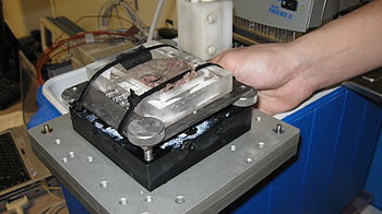
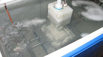
The methods of this work involved the measurement of temperature and synchronous ultrasonic imaging of tissue in a water bath. The tissue was usually a piece of turkey purchased from a local grocery store or a piece of preserved rabbit liver. The tissue was placed in experimental housing as seen in Figure 1. This housing was then placed in a water bath in an insulated container, as seen in Figure 2. There are four main components to the experiment, all of which interface with each other via a MATLAB script. The components include: 1) the DATAQ, which acquires temperature data, 2) the ThermoHaake, heats and circulates the water bath, 3) the ultrasound transducer, which acquires ultrasonic images of the tissue, and 4) the Newport, which is a mechanical actuator which moves the ultrasound transducer so it can take pictures of multiple slices of tissue. The DATAQ, ThermoHaake, and Newport are all named after the makes or models of the device.
Contents
The DATAQ and Temperature Data Acquisition
Seen in Figure 2 are thermocouples inserted into the tissue. A clearer view of the thermocouples can be seen in Figure 3. These thermocouple attached to a DATAQ device, seen in Figure 4. This device is used to acquire temperature data from multiple (up to 8) channels via thermocouple. This acquisition is done via interface with a computer program called Windaq. Windaq produces plots of temperature versus time for the given channels of the DATAQ. MATLAB interfaces with Windaq which interfaces with the DATAQ. This is the path that the flow of temperature data takes.
The ThermoHaake, Water Heating, and Water Circulation
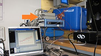
A second device used in the experiment is the ThermoHaake (the device with the screen as seen in Figure 5). This device has 3 purposes: 1) Heat the water bath, 2) Circulate the water bath, and 3) Measure the temperature of the water bath.
The water bath is heated by heating coils on the underside of the ThermoHaake. This heating system is used to bring the water temperature from 36 degrees Celsius to 70 degrees Celsius.
The water bath is circulated by a pumping filtration system. In the same complex as the heating coils are an incoming and an outgoing pump. This system takes up water from the bath, filters the water, and then deposits the water back into the bath. This mechanism circulates the bath such that the water is in constant motion. By circulating the water, the ensures that the water bath heating will be very nearly uniform.
The water bath temperature is measured by a third component of the complex under the ThermoHaake. This device is a thermistor that measure the sample of water below the ThermoHaake, but this sample is treated as representative of the entire water bath, especially because of the circulation. The thermistor is also treated as the "truth" for temperature measurement, that is to say that it is treated as having zero error in its measurement.
This system measures the temperature in the tissue from 4 different channels and at the same time measures temperature from two points in the water bath. The two points outside of the tissue are the space directly beneath the ThermoHaake and the space on the opposite end of the insulated container. These temperature measurements are to observe any temperature gradient in the water.
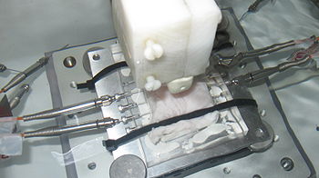
Figure 3 - The housing placed in the water bath with a clear view of the thermocouples
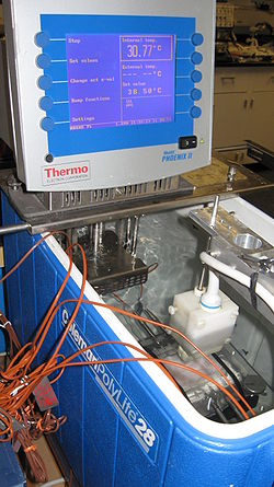
Figure 5 - Overview of the Experimental Setup
The Ultrasound Transducer
The ultrasound transducer is used to take images of the tissue in the area of the four thermocouples in the tissue. In the end, the research this experiment is contributing to will find the correlation between these images and the temperature measurements in the tissue at the time of the image. The transducer interfaces with a computer program called Terason. Terason is a user interface that allows for easy use of the transducer. MATLAB calls the Terason to open and take ultrasonic images of the tissue. This images are saved in a directory on the computer.
The Newport
The Newport
